The Amazing Eye: The Mechanics of Vision
Two parts comprise the visual system: The optical system (your eye) and the perceptual system (your brain). Fundamentally, the brain analyzes the information your eye gathers.

Sight is an integral part of our lives—from the moment we awaken to the moment we fall asleep, we consume information visually. Vision is a complex sense. We constantly rely on our eyes to understand the world around us, and our visual system processes millions of pieces of information every second.
Have you ever wondered about the process behind our eyes and brains that enables us to see? Understanding basic eye anatomy and the mechanics of the visual system can help us grasp things that cause vision problems and conditions.
Eye Anatomy
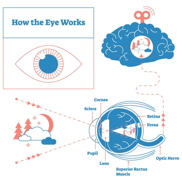
Understanding the mechanics of the eye helps to understand basic eye anatomy. The eye functions like a camera with a lens, film, and lens cover. The main parts of the eye include:
Cornea
The cornea is the clear protective coating on the front of the eye that allows light to pass through it without distortion. It covers the colored iris. The eye lens processes the images transmitted through the cornea to the retina.
Conjunctiva
The conjunctiva is the clear covering (like cellophane) of the sclera, the white part of the eye. When it gets irritated or infected, it becomes red, called conjunctivitis.
Eyelids
Eyelids play a vital role in preserving sight. They keep moisture inside, so our eyes don’t dry out from exposure to air and foreign particles outside of the eye.
Lacrimal System
The lacrimal (tear) system is a miniature drainage network that prevents tears from rolling down cheeks. From the eye’s surface, tears flow along the edge of the lids toward the nose. Before reaching the corner of the eye, the tears slide into two tiny drain tubes and then into a larger tube, which carries the tears into the nose.
Lens
The lens lies directly behind the pupil in a sac-like capsule and is responsible for focusing the picture. Your lens is usually clear and transparent. It focuses images onto the retina, which acts as the film recording the picture. The optic nerve then transmits the image to the brain, and the brain interprets it. It’s the brain that does the actual seeing.
Macula
The macula is a part of the eye that contains special light-sensitive cells to see fine details. It is located in the retina. The macula is an integral part of the eye because even small changes can cause severe vision loss.
Optic Nerve
The optic nerve is the pathway connecting the eye to the brain. The nerve fibers carry messages from the eyes to the brain.
Orbit
The eyeball sits in the orbit, a bony housing that protects the eyeball on all sides, except in the front, where the lids protect it. The orbital contents—fat, muscle, blood vessels, and glands—are located between the bony housing and the eyeball.
Pupil
The dark center of the iris (the colored part of the eye) is called the pupil. The pupil determines how much light is needed for the eye to see correctly and changes sizes to adjust for changes in light.
Retina
The retina is the light-sensitive part of the eye. It has one major artery and one major vein called the central retinal vein. An area on the retina, called the optic disc, is where all the nerve fibers join to become the optic nerve, which connects to the brain.
Sclera
The sclera is the “white” part of the eye. It’s made of fibrous tissue and protects the inner-eye structures.
Vitreous
The vitreous is the clear jelly-like substance that fills the middle part of the eye.
The Mechanics of the Visual System
Two parts comprise the visual system: The optical system (your eye) and the perceptual system (your brain). Fundamentally, the brain analyzes the information your eye gathers.
The Optical System
A simple explanation is that light travels through our eyes, where optic nerves process this information and send signals to the brain. But a deeper dive into this process demonstrates the unique and complicated system that allows us to see.
First, light travels through your cornea — the clear, dome-shaped protective layer at the front of your eye, which can bend and refract light. Next, the light passes through your pupil. Then, depending on the amount of light reflected into the eye, the iris changes in size to allow more or less light. For example, if you step outside on a bright day, the light might feel blinding until your eyes adjust.
Once the light enters the eye, it focuses on the retina. The lenses change shape depending on the distance of the object you’re viewing. It’s called accommodation and involves ligaments surrounding your eye that pull or release the lens to focus correctly. The action doesn’t happen perfectly for people who are nearsighted or farsighted.
Once the light focuses on the retina, it’s transformed into electrical impulses. Then, it begins a journey, traveling from the optic nerve to the brain.
The Perceptual System
Our eyes capture light from objects around us, sending the information to our brain. Our eyes don’t actually “see” anything. The visual cortex does that. The information from the impulses that travel from the optic nerve to the brain delivers an upside-down image. The brain then transforms the image right side up so we can comprehend it.
The Effects of Glaucoma
The front part of the eye is filled with a clear fluid (called aqueous humor) made by the ciliary body. The fluid flows out through the pupil. It then reaches the eye’s drainage system, including the trabecular meshwork and a network of canals. The inner pressure of the eye (intraocular pressure or “IOP”) depends on the balance between how much fluid is made and how much drains out of the eye.
In most glaucoma cases, fluid builds up because the eye’s drainage system becomes blocked, so the intraocular fluid cannot drain. The buildup of fluid causes pressure inside the eye, damaging sensitive nerve fibers. If the pressure remains too high, the additional pressure on the sensitive optic disc can lead to permanent vision loss.
Although there is no cure for glaucoma yet, the key to preventing vision loss from glaucoma is early detection, diagnosis, and treatment.
Everyone is at risk for glaucoma, but certain groups and circumstances are associated with greater risk. In addition, there can be several contributing causes of glaucoma. Talk to your eye doctor about your chance of getting glaucoma, especially if you have one or more of these risk factors.
Help Us Keep Eyes Healthy
The diligent work of researchers continues to lead to a better understanding of glaucoma every day. As a result, there’s great hope for new and improved treatments, including superior drug delivery methods, laser treatments, and less invasive surgical techniques. You can help make that happen!
Every contribution helps bring us closer to finding a cure for glaucoma. Whether you donate cash or stock, create a fundraising event, or even donate a vehicle or boat, your donation will give hope to those living with glaucoma and accelerate our research.
Posted on April 6, 2022.