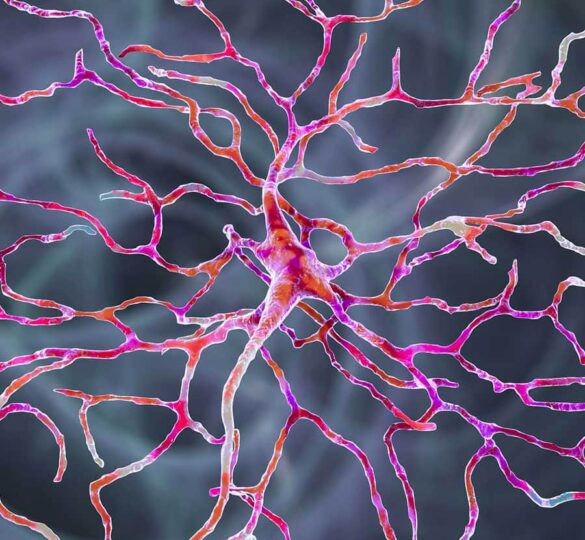
Protecting Retinal Ganglion Cells
This article summarizes published findings from Catalyst for a Cure (CFC1) research scientists and subsequent discoveries by Dr. Rebecca Sappington's lab at Vanderbilt and Wake Forest.

The retina is a thin tissue in the back of the eye containing photoreceptor nerve cells. These nerve cells, known as retinal ganglion cells (RGCs), change the light rays that enter the eye into electrical impulses and send them through the optic nerve to the brain where images are perceived.
The cells that comprise the retina and the brain can be divided into two main classes, neurons and glial cells. RGCs are neurons and glial cells are non-light sensing cells that support them.
Early work by Catalyst for a Cure (CFC) research scientists examined both neurons and glial cells in glaucoma. Neurons and glial cells communicate with one another via signaling proteins that are released by one cell and received by another. Cytokines are one such family of signaling proteins that can act to both increase and decrease the survival of neurons.
Research Findings
Our early work, funded by the CFC, identified the cytokine interleukin-6 (IL-6) as a potential protector of retinal ganglion cells in glaucoma. We found that IL-6 released by microglia, the smallest glial cell, can protect RGCs from pressure-induced death in a dish (Sappington et al., 2006).
Inspired by these early CFC findings, we established an independent research program to determine how and why IL-6 protects RGCs in glaucoma.
We found that the role of IL-6 in glaucoma was much more complex in an intact visual system. In our studies, removal of IL-6 resulted in preservation of RGCs, but did not alter all indicators of glaucoma damage in the optic nerve (Echevarria et al., 2017).
The difference between IL-6 activities in the intact system and the isolated system likely arise from another surprising finding – IL-6 is also important for the development and structure of RGCs in healthy optic nerve.
We found that healthy RGCs use IL-6 to maintain microtubules. Microtubules are the skeleton of RGCs in the optic nerve that allow communication between the RGC body in the retina and the feet (or terminals) in the brain. We found that IL-6 builds and maintains a straight and stable microtubule skeleton that is necessary for communication within RGCs (Wareham and Echevarria et al., 2021).
Interestingly, this microtubule mechanism is also shared by neurons in the spinal cord, suggesting that it is important for stabilizing neurons that span long distances to deliver messages to and from the brain. Remarkably, this microtubule mechanism is also linked to cancer. Many chemotherapy drugs shrink tumors by destroying microtubules. Resistance to chemotherapy is associated with preservation of microtubules via this mechanism in several types of cancer. This raises the possibility that cancer cells preserve themselves by tapping into a mechanism that is designed to stabilize RGCs and other “long distance” neurons!
The Big Picture
The complexity of IL-6 activities in glaucoma is likely due to both a loss of healthy IL-6 signaling and an increase in disease IL-6 signaling. As illustrated by our IL-6 findings, it is critically important to understanding how healthy and disease signaling intersect to not only treat glaucoma damage in RGCs, but to also prevent glaucoma damage in healthy RGCs.
This article summarizes published findings in the:
- July, 2006 edition of Investigative Ophthalmology & Visual Science: “Interleukin-6 Protects Retinal Ganglion Cells from Pressure-induced Death” by Rebecca Sappington, Matilda Chan, and David Calkins.
- May, 2017 edition of Frontiers in Neuroscience: “Interleukin-6 Deficiency Attenuates Retinal Ganglion Cell Axonopathy and Glaucoma-Related Vision Loss” by Franklin Echevarria, Cathryn Formichella, and Rebecca Sappington
- October, 2021 edition of iScience: “Interleukin-6 Promotes Microtubule Stability inAxons via Stat3 Protein–protein Interactions” by Lauren Wareham, Franklin Echevarria, Jen Sousa, Danielle Konlian, Gabrielle Dallas, Cathryn Formichella, Priya Sankaran, Peter Goralski, Jenna Gustafson, and Rebecca Sappington
First posted September 11, 2006; reviewed and updated by Rebecca Sappington, PhD, August 2, 2022

Rebecca M. Sappington, PhD
Rebecca M. Sappington, PhD is an Associate Professor of Neurobiology and Anatomy and Associate Professor and Vice Chair for Research in Ophthalmology at the Wake Forest University School of Medicine in Winston-Salem, North Carolina.