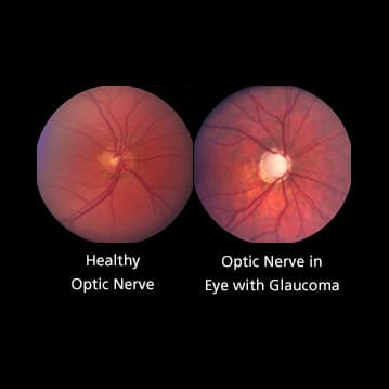
Optic Nerve Cupping
Both people with and without optic nerve damage have optic nerve cupping, although those with glaucoma tend to have a greater cup-to-disc ratio.

The optic nerve carries impulses for sight from the retina in the eye to the brain. It is composed of millions of retinal nerve fibers that bundle together and exit to the brain through the optic disc located at the back of the eye. The optic disc has a center portion called the “cup” which is normally quite small in comparison to the entire optic disc.
In people with glaucoma damage, because of increased pressure in the eye and/or loss of blood flow to the optic nerve, these nerve fibers begin to die. This causes the cup to become larger in comparison to the optic disc, since the support structure is not there. Optic nerve cupping progresses as the cup becomes larger in comparison to the optic disc.
Both people with and without optic nerve damage have optic nerve cupping, although those with glaucoma tend to have a greater cup-to-disc ratio. A cup to disc ratio greater than six-tenths is generally considered to be suspicious for glaucoma.
Through periodic photographs of the optic nerve, the ratio of the cup to the disc can be monitored. This helps the doctor determine whether or not damage is still occurring to the nerve fibers with current treatment and/or if treatment should be modified.
Article by John S. Cohen, MD and Harry A. Quigley, MD. Last reviewed March 16, 2022.
John S. Cohen, MD is Director Emeritus of the Glaucoma Service at the Cincinnati Eye Institute (CEI). He is a founding member of the American Glaucoma Society, and a recipient of the Honor Award and Senior Honor Award from the American Academy of Ophthalmology.