Gonioscopy: What Is It And Why Is It Needed?
Part of a complete glaucoma eye exam, gonioscopy is a test performed by an eye doctor to evaluate the internal drainage system of the eye.
Gonioscopy: What Is It?
Gonioscopy is performed during the eye exam to evaluate the internal drainage system of the eye, also referred to as the anterior chamber angle. The “angle” is where the cornea and the iris meet. This is the location where fluid inside the eye (aqueous humor) drains out of the eye and into the venous system.
Under normal circumstances, the angle cannot be seen on exam. A special contact lens prism placed on the surface of the eye allows visualization of the angle and drainage system.
Why Is It Needed?
The pressure inside the eye is maintained by constant production and drainage of fluid. If the drainage system is not working properly, the pressure inside the eye, also known as intraocular pressure, can increase. High intraocular pressure can cause damage to the optic nerve, the “cable” that sends images from the eye to the brain. This kind of damage is called glaucoma, the second leading cause of blindness worldwide.
By looking at the “angle,” physicians can determine if it is open or closed as well as if there are abnormal blood vessels, adhesions (synechiae), or damage from previous eye trauma. A closed angle is an abnormality that can predispose the patient to have a sudden or rapid increase in intraocular pressure. This increase in pressure can cause a very serious, acute form of glaucoma that can be treated and even prevented with laser treatment (iridotomy) if the predisposing angle abnormality is recognized using gonioscopy.
In addition, gonioscopy allows the eye doctor to note more subtle characteristics of the eye’s drainage system, in order to guide his or her diagnosis and treatment plan.
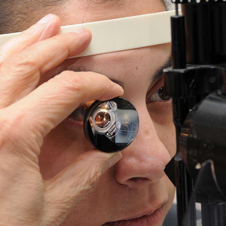
How Is It Done?
Gonioscopy is performed with the head positioned in the slit lamp (the special microscope used to look at the eyes). After numbing the eye with drops, a special contact lens is placed directly on the eye and a beam of light is used to illuminate the angle. While the eyelids may feel the presence of the lens, there is typically no pain associated with this exam. Examination of both eyes typically takes a few minutes.
Article by Kathryn E. Bollinger, MD and Dr. Michael Westafer. Last reviewed on March 8, 2022.

Kathryn E. Bollinger, MD, PhD
Kathryn E. Bollinger, MD is a glaucoma specialist, Associate Professor of Ophthalmology, and Director of Glaucoma Service at the Medical College of Georgia at Augusta University. Dr. Bollinger completed her ophthalmology residency and glaucoma fellowship at the Cole Eye Institute, Cleveland Clinic.