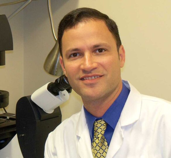
Biomarkers and Drug Discovery: Jeffrey L. Goldberg, MD, PhD
Dr. Goldberg talks about the Catalyst for a Biomarker Imitative and its impact on glaucoma research.

Jeffrey L. Goldberg, MD, PhD presented research advances from the Catalyst for a Cure biomarkers team at the Glaucoma 360 New Horizons Forum on February 3, 2017 in San Francisco.
The title of Dr. Goldberg’s talk was “Biomarkers and Drug Discovery: Integrating therapeutic and diagnostic models.”
Video Transcript
Dr. Jeff Goldberg: I really want to thank the Glaucoma Research Foundation. Obviously, what we’re going to talk about today would not come to fruition without the support of the Glaucoma Research Foundation and of course, all of their supporters.
So, we’ve heard today about the unmet needs in glaucoma. We need better intraocular pressure lowering approaches: pharmacologic or surgical. Of course, my laboratory, we’re very focused on the treatments that we need that go beyond interocular pressure: neuroprotection, regeneration, neuroenhancement.
And closely related to that need is the need for better biomarkers which can improve the measures of risk, the diagnosis, the progression, and importantly, as we will come to by the end of the talk, serve as an important opportunity to speed clinical trials of therapeutic candidates.
So, that’s where [we see] this impact of the Glaucoma Research Foundation. We’ve really felt it personally, seeding these innovate Shaffer grants that David just introduced. This Catalyst for a Cure program, and now this Catalyst for a Biomarker Imitative. Now, in this we’re very focused not on intraocular pressure at all but on the back of the eye, on the retinal ganglion cells. Of course, the retinal ganglion cells and their axons are what degenerate in this disease. There’s no retinal ganglion cell regeneration or replacement after optic nerve injury.
So, I want you to pause a moment and imagine (here’s my laboratory group, who I’m very grateful to, pausing and imagining themselves, on their iPhones etcetera). So, pause for a moment and imagine, how do we diagnose glaucoma? We all are aware of the difficult that we have doing that. Visual field testing, current OCT technology. How do we know if we’re being treated properly, adequately? The patient sitting in front of you, are they on enough treatment? Of course we’re going to have to wait a couple of years and see whether their visual field gets worse in order to look back and say whether they were being adequately treated. Obviously, that’s not the ideal. And then how do we develop and test new therapies?
Now this latter element, it turns out that we have a real opportunity because as Dave Calkins and others have now demonstrated very beautifully in animal models, and we even have very good data now in humans; in glaucoma there’s an injury that happens first, and it’s the death or loss of the cell that happens later in the disease. So, intraocular pressure may be elevated, axon transport fails, the axon ends up being physically damaged; the retinal ganglion cells die relatively late in the disease.
So how do we measure glaucoma before it’s too late? How do we intervene in that window of opportunity? So right now, we have a number of ways that we measure glaucoma. Of course, visual field testing, looking at the optic nerve, optic nerve atrophy, nerve fiber layer thinning, using optical coherence tomography, but the real question is can we back that up significantly and be able to tell whether the patient sitting right there in the chair is in trouble? To that we’ve really been focusing on whether there’s been a loss of metabolic function or specific features in retinal ganglion cells and their processes.
So again, we’ve already introduced the Catalyst for a Cure. These are my collaborators and I’m going to show work from all of our labs. Alf Dubra, Medical College of Wisconsin who is recently recruited to Stanford University. Myself, a glaucoma specialist and neuroscientist. I was at UC San Diego. About a year and a half ago I moved to Stanford University. Andy Huberman, one of the really premier neuroscientists studying the visual system was at UCSD, now at Stanford University. And Vivek Srinivasan, an optical engineer very focused on how we image the eyes, who was at Harvard/MGH and came almost to Stanford University but is thankfully right down the street at UC Davis. So, coincidentally we’ve all converged here in the Bay Area.
So, let me just lay out the principle. The principle has been team science; and the principle has been to take the neuroscientists, glaucoma specialists, put together with the optical engineers, let’s discover some fundamental biology, and then implement innovative engineering as a way to push that out of the laboratory and into the clinic. I’ll just highlight a couple of examples.
So, one example comes from the study of the retinal ganglion cells degenerating in glaucoma. Andy Huberman’s group published last year that retinal ganglion cells, you can classify them in a number of different ways. One way you can classify them is the ganglion cells that fire when lights turn on and the ganglion cells that fire when lights turn off. Those are understandably called the ‘on’ retinal ganglion cells and ‘off’ retinal ganglion called. What he and his group discovered was that actually the ‘on’ retinal ganglion cell dendrites were not easily impacted in glaucoma models. Their dendrites did not retract very readily. But the ‘off’ retinal ganglion cell dendrites retracted very early in the disease.
So, with that in mind, with this new understanding of fundamental biology, the group went back into the engineering side and said well, what can we do to image ‘off’ retinal ganglion cell layers of the inner plexiform layer, and also to design a new visual field exam that could measure the ‘off’ retinal ganglion cells separately from the ‘on’ retinal ganglion cells? Neither of these have really been done effectively up to this date.
Let me give you another example. We discovered in the laboratory, our lab and others, that mitochondria — these are the little energy powerhouses inside the cells, inside all cells including retinal ganglion cells — they fragment and stop moving in retinal ganglion cell axons very early in glaucomatous insults. Now, mitochondria are already very tightly associated to neural degeneration in the visual system and throughout the central nervous system. So, there is a very good indication to go after that.
It turns out that after optic nerve injury the mitochondria decay very rapidly. They decay in the retina not just at the site of injury in the optic nerve, but back in the retina where we can be imagining them. And in fact, we can image these axon mitochondrial dynamics both in the optic nerve and in the retina in animals. So importantly we again turn to our engineering colleagues who are asking us what should we measure? We said well, another great thing to measure might be the mitochondria structure and function.
And so, both Alf and Vivek have put together approaches using OCT and adaptive optics that we’re now merging into uniform instrumentation where we can really do metabolic imaging live, non-invasively, in the retina of our patients with glaucoma. We’re able to measure the mitochondria moving. If you look on the video to your right those are mitochondria that are moving down retinal ganglion cell axons. (That’s a little video that’s looping in real-time, a few seconds at a time.)
And Alf Dubra is now using adaptive optics to image what we think are the same mitochondria moving in retinal ganglion cell axons. (These little kind of wormy contortions, if you will, that’s a good scientific description.) Those are what we think are mitochondria again slivering in and out of the retinal ganglion cell axons. So, we’re now in a position to ask whether those retinal ganglion cell axons and those mitochondria — putative mitochondria — are fragmenting in response to interaction or pressure elevation? Are they predictive of the poor health of a glaucoma patient?, etcetera.
Let me just give you one more example of how we’re taking fundamental discovery out of the laboratory and moving it into the clinic. There we’re returning to the therapeutic side and finding exciting advances in optic nerve regeneration and retinal ganglion cell transplantation. Sort of addressing the stem cell question, if you will.
Andy Huberman published a really beautiful paper this past year where what they did is they actually combined some molecular therapeutic manipulations of retinal ganglion cells with visual stimulation. The kind of visual stimulation that we could design and give to our patients. What his group found is that not only does that promote axon regeneration all the way back down the nerve, but in fact those axons go back to the right areas that they’re supposed to get to in the brain — the right brain nuclei — and restore some measures of visual function.
Similarly, in the laboratory we’ve been studying cell replacement therapy. Can we transplant retinal ganglion cells from one animal to another thinking about eventually doing that in people? For this we inject themselves into the vitreous, into the center of the eye, and what we’re finding is that a subset of the injected retinal ganglion cells can go into the retina, the green cells are the donor cells, they extend all of their axons and dendrites. Those are the dendrites. That’s the dendritic tree where they’re supposed to be collecting all that visual information. In fact, when we flash lights at the retina, the black bars here are the light flashing on the retina, and these donor retinal ganglion cells are responding to those light bars. So, they’re integrating electrophysiological into the retina. They’re showing the various different types of physiology inside the retina that we expect them to have. Not only that, their axons, a subset of them, their axons are going all the way down the optic nerve. Here the axons going down the optic nerve, across the optic chiasm, and into the expected targets in the brain: the lateral geniculate and the superior colliculus.
So again, we’re taking these exciting advances in optic nerve regeneration and retinal ganglion cell transplantation and asking how do we translate these into the clinic and how do we use biomarkers effectively? So, we’re now designing clinical trials to test visual stimulation as a way to promote vision restoration in humans.
We’re moving therapeutic candidates from the lab to the clinic. And importantly, we’re combining therapeutic trials with testing these new biomarkers out of the laboratory, in humans, to ask if the biomarkers can let us detect disease or improvement even faster.
So, we’re implementing these tools. We’re bridging to human testing. Testing these structural and functional markers in glaucoma suspects and patients and importantly, in patients enrolled in clinical trials for vision restoration, which I know are few and far between. But let me finally, in the last couple of minutes, introduce that.
Now we’ve heard a lot today about how glaucoma damage takes a long time to measure. So, it’s a slow disease on average and if we’re going to bend that green curve with a therapeutic, maybe try to bend it up to that orange curve, that can be tough. But if we pick therapeutic candidates that might enhance function and show us an acute improvement in vision, or use a metabolic biomarker that again gives us the hint that we’ve acutely improved the health of retinal ganglion cells, that can give us confidence in a short period of time that the therapeutic candidate we are studying has promise.
So, let me give an example. We undertook, a couple of years ago, a phase one trial for one such neuro enhancing therapeutic called ciliary neurotrophic factor or CNTF. We tested it in glaucoma. This was delivered through an implant made by a company called Neurotech. It’s a semi-permeable membrane filled with cells that are secreting the CNTF into the eye over the long term and it’s inserted through the eye. (I won’t show you what the whole surgical video.) Afterwards, it’s implanted in the eye and it just stays inside the eye where it can secrete and feed the retina and the optic nerve. We recruited 11 glaucoma patients and of course our primary outcomes through that first trial with safety. There were no serious adverse events. Also, no effect on intraocular pressure. So, that gave us great confidence in moving forward in glaucoma patients.
Importantly we also looked for signals of efficacy and in these patients although, we can’t do statistics, we saw improved visual field indices in the treated eye compared to the untreated eye. We saw thickening of the nerve fiber layer. So, we have correlated structure function improvements and importantly this was not just a big effect in a couple of patients. It was almost every patient who showed these very similar changes. So, it had a very consistent biological effect on retinal ganglion cells and vision. So, again, our interim conclusions were that there is a suggestion certainly of biological activity. That these neurotropic factors could promote survival and regeneration and that has led us know to phase two evaluation.
It is in this phase two evaluation where patients are now randomized to either sham surgery or getting the CNTF implant. Although the ones who get the sham have a chance to get crossed over to get the real thing a year later. We’re using this advanced biomarker imaging endpoints as part of our way to detect whether the therapeutic is having a positive effect early in the trial. So, the patient recruitment has started already. We’re recruiting patients with a range of visual field defects. The primary endpoint is in months, not in years; first data will be here by late summer.
So, in summary, we are thinking about translation, we’re thinking about neuroprotection, regeneration, neuroenhancement in glaucoma. How do we improve visual function? And we’re obviously very focused on the biomarkers that we think are really going to be required to measure these, to accelerate the development testing of candidate therapies. So, I’ll stop there, and thank you very much.
(End Transcript.)
Posted on March 22, 2017; Reviewed on April 20, 2022