Catalyst for a Cure 2018 Research Progress Report
Biomarkers can provide physicians with better tools to diagnose and manage glaucoma before vision is lost, as well as accelerate better treatments for glaucoma.

The Catalyst for a Cure (CFC2) Biomarker scientists are now evaluating their novel biomarkers in glaucoma patients. The newly identified markers of glaucoma can be detected with unique imaging devices and diagnostic tests developed by the team.
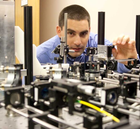
The imaging devices include adaptive optics scanning laser ophthalmoscopy (AOSLO), specifically developed to provide extremely high resolution images of the retina, and an optical coherence tomography (OCT) ophthalmoscope utilizing visible light to image specific layers in the retina with unprecedented depth resolution and contrast. The diagnostic test is a physiologic measurement probing retinal ganglion cell subtypes to assess visual functions that may be lost early in glaucoma.
The high resolution AOSLO images make it possible to see retinal ganglion cells directly and to visualize the transport of mitochondria within the cells. It is hypothesized that changes in the shape or motion of the mitochondria will predict glaucoma and/or glaucoma progression. The visible light OCT images provide information on the thicknesses of specific layers in the retina, which may change early in glaucoma before vision loss occurs. The system can also yield information on the oxygen being consumed by these cells, which may relate to their health. Early measurements on glaucoma patients and normal eyes are helping to refine the instrumentation and determine the sensitivity and specificity of each biomarker for detecting glaucoma.
Through their earlier research, the CFC scientists also discovered that certain retinal ganglion cells were more susceptible to damage from glaucoma than others. These cells were hypothesized to be the “canary in the coal mine” that might warn of glaucoma in the earliest stages of disease before vision loss is severe enough to be detected by conventional visual field tests. The team designed a new test with patterns of light and dark spots of light, to specifically assess these “canary” subtypes of retinal ganglion cells. This test is more objective and might be more sensitive for glaucoma than a conventional visual field test. It is also much easier for patients to take compared to conventional visual fields, because it does not demand as much attention. The team is now testing both glaucoma patients and normal individuals with an optimized test to determine possible correlations with the stage of the glaucoma and even with the impact of therapy to restore vision.
The CFC team is testing a number of hypotheses regarding the biomarkers they have identified and their ability to both detect and monitor glaucoma. Glaucoma Research Foundation is continuing to fund the CFC Biomarker scientists for another year so they can demonstrate positive correlations between the health of the retinal ganglion cells and the particular measurements they are performing. Once these relationships are defined, the hope is that NEI and other funding, and ultimately collaborations with industry partners, will result in new clinical instruments that will benefit many more patients with earlier diagnoses and more sensitive measures of changes resulting from glaucoma treatments.
2018 Research Progress Video:
First posted on April 2, 2018; Reviewed on May 20, 2022