Why Retinal Ganglion Cells Are Important in Glaucoma
Retinal ganglion cells (RGCs) are particularly important in glaucoma because they are the cells that are damaged by the disease.

The retina is a thin tissue in the back of the eye containing different kinds of nerve cells. Among these are retinal ganglion cells (RGCs) — and they are particularly important in glaucoma because they are the cells that are damaged primarily by the disease.
Retinal ganglion cells process visual information that begins as light entering the eye and transmit it to the brain via their axons, which are long fibers that make up the optic nerve.
There are over a million retinal ganglion cells in the human retina, and they allow you to see as they send the image to your brain. Once RGCs die in glaucoma, they are not replaced. Unlike peripheral nerve cells in other parts of the body, RGCs are part of the body’s central nervous system, which does not regenerate once damaged.
Researchers are still studying how and why certain nerve cells lose their ability to function, or in some cases “turn themselves off.” Doctors and scientists know that increased intraocular pressure (IOP) has a detrimental effect, and that lowering IOP can help preserve vision in glaucoma patients. They also know that preserving the retinal ganglion cells and their axons is necessary for sight.
According to Harry A. Quigley, MD (Wilmer Eye Institute, Johns Hopkins Medicine), “Ganglion cells are damaged by prolonged eye wall stress and this is the cause of damage to your vision in glaucoma. This means that the higher the pressure, the greater the chance for glaucoma. However, not everyone’s eyes react to pressure in the same way.”
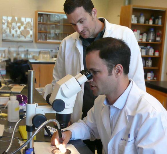
Jeffrey L. Goldberg, MD, PhD, a member of the Catalyst for a Cure (CFC2) Biomarkers research team, notes, “because the retinal ganglion cell axon stretches from the retina through the optic nerve to the brain, its projections also become damaged by glaucoma. In addition to treatments directed at lowering eye pressure, still the mainstay of glaucoma therapy, there may be opportunities to develop new types of treatments directed at the retina and the brain.”
Andrew D. Huberman, PhD, another CFC Biomarkers Team principal investigator, adds his take on RGCs in glaucoma: “My lab is investigating the biology of the ganglion cells, to examine which ones are vulnerable early in glaucoma and how best to treat them on the molecular level to keep them healthy and prevent them from dying.” Dr. Huberman is enthusiastic about the potential for helping damaged RGCs to recover. “Eventually, we hope to even repair or regenerate some of the visual system if it is already damaged,” he said.
Article reviewed by David J. Calkins, PhD.
Posted on January 18, 2018; Reviewed on March 23, 2022

David J. Calkins, PhD
Dr. Calkins is the Denis M. O’Day Professor of Ophthalmology and Visual Sciences and Vice-Chairman and Director for Research at The Vanderbilt Eye Institute, Vanderbilt University Medical Center in Nashville, Tennessee.