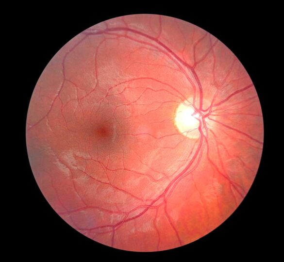
Imaging of the Optic Nerve: What is it and why is it needed?
Glaucoma specialists take pictures of the optic nerve in order to measure damage to this important cable that connects your eye to your brain.

There are several types of pictures, and each one provides different information about the amount of optic nerve tissue lost as well as the rate of nerve fiber thinning or “progression” that occurs when glaucoma is not controlled.
The goal of today’s glaucoma therapy of lowering intraocular pressure is to slow progression, and imaging tests are an important tool for monitoring progression. Imaging of the optic nerve is complementary to visual field testing and using both together is more useful than each test alone.
This is because imaging records the anatomy, or structural features of the eye, while visual fields assess what someone actually sees, or the function of the eye. In general, optic nerve imaging is more useful in glaucoma suspect and early-to-moderate glaucomatous patients rather than advanced disease.
Optic Nerve Photos and Scans
Two common imaging tests include a simple high-resolution color photograph with a very bright flash from a professional camera, and a quick laser scan of the optic nerve. Scans can detect small nerve fiber layer changes of the optic nerve at the micron level.
Viewing the optic nerve through lenses and a slit lamp is the best way for your doctor to assess the optic nerve for glaucoma. Your doctor may document this assessment either with drawings or with optic disc photos. A color photograph provides a more accurate baseline for future comparison. Photographs do not get outdated, and the information contained in them can be very useful for demonstrating “no change” years later.
Compared with a photograph, optic nerve scans contains more objective, quantitative information for staging of disease progress. Scans may also be better for determining when glaucoma is getting worse. However, every few years, the software is updated, and every decade the technology advances dramatically just like computers. This sometimes makes the data less useful because it can be difficult to compare new versions with old ones.
Since optic nerve damage cannot be reversed by any current therapy, close surveillance with structural as well as functional testing, combined with early intervention while balancing the risk of side effects, is key to keeping glaucoma under control and to preserving sight.
Types of Optic Nerve Scanning Technologies
- OCT – Optical Coherence Tomography (Devices include: Cirrus HD-OCT, RTVue-100, Spectralis, Topcon 3D-OCT 2000, and others)
- CSLO – Confocal Scanning Laser Ophthalmoscopy (HRT)
- SLP – Scanning Laser Polarimetry (GDx)
OCT (Optical Coherence Tomography) Video
Article by Robert Chang, MD. Last reviewed March 28, 2024.

Dr. Chang completed his ophthalmology training at Washington University in St. Louis and then completed a research and clinical fellowship at the Bascom Palmer Eye Institute at the University of Miami. He currently is an Assistant Professor at Stanford University at the Byers Eye Institute in Palo Alto, California.