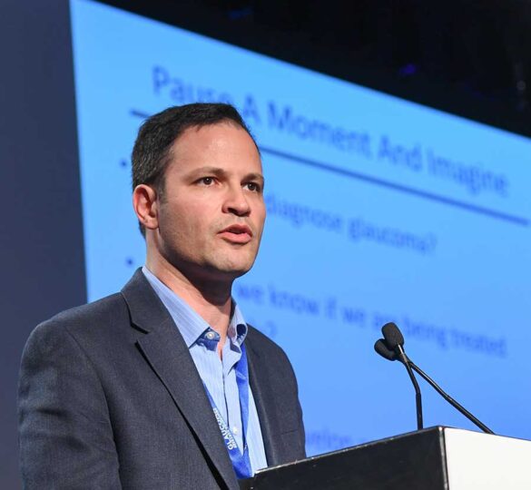
2018 Weston Lecture by Jeffrey Goldberg, MD, PhD
Listen to the 2018 Weston Glaucoma Research Lecture delivered by Jeffrey Goldberg, MD, PhD, a principal investigator in the Catalyst for a Cure.

“A Clear Path to Vision Restoration” — On October 16, 2018 Jeffrey Goldberg, MD, PhD delivered the Weston Glaucoma Research Lecture in Palo Alto, Calif., presented by Glaucoma Research Foundation.
Click the link below to listen to the 30-minute lecture:
About the Lecture
Jeffrey L. Goldberg, MD, PhD is a principal investigator in the Catalyst for a Cure Biomarkers Team funded by Glaucoma Research Foundation in San Francisco, CA. Catalyst for a Cure brings together scientists from different backgrounds to work collaboratively to understand glaucoma and find ways to improve treatment and ultimately cure this blinding disease. Dr. Goldberg’s research is directed at neuroprotection and regeneration of retinal ganglion cells and other retinal neurons. His laboratory at Stanford University School of Medicine is developing novel stem cell and nanotherapeutics approaches for ocular repair, studying retinal ganglion cell development, survival and axon regeneration in glaucoma, and investigating the cellular basis for the developmental loss of axon growth ability.
Audio Transcript
Dr. Jeffrey Goldberg: Thank you very much for the wonderful opportunity. It’s really great to be here. The last time when I came to this some years ago, it was the previous venue, and obviously this is a beautiful venue and a wonderful place that is able to host these wonderful events.
My thanks- I remember meeting the Weston family some years ago, and so I got a chance to say hi to you earlier and hello again now, and so just thank you very much for having this great event.
I was asked to talk about the present and particularly the future of glaucoma diagnosis and treatment, and I know a lot of familiar faces, Glaucoma Research Foundation, supporters, glaucoma patients in the audience. I know we all struggle with the present of glaucoma treatment, and that’s why I thought it’d be really tantalizing to talk a bit about the future of glaucoma diagnosis and treatment and how we’ve made such great progress, in particular under the auspices of the Glaucoma Research Foundation over these last years.
The Glaucoma Research Foundation, as many of you know, seed innovative ideas, new ideas, through their Schaffer Grants and other grants, programs for innovative research, and have a real flagship program in this catalyst for a cure, which over its first decade focused on understanding the underlying causes, what we call pathophysiology of glaucoma, and most recently in the group that I participated in collaborative, figuring out better ways to measure the disease, and why do we need to measure the disease better? So we can test new potential cures for the disease, new ways to restore vision, and that’s what I’m going to talk about today and what the focus is for these coming years.
I’ll acknowledge up front that I’ve had great collaborators throughout these years, and I’ll mention a few of their names along the way. I’m also enormously fortunate to have settled three years ago at the Byers Eye Institute at Stanford. It’s a wonderful department. I know we get to see some of your eyes there. It’s full spectrum care, and it’s just been a really wonderful, collaborative, innovative area with lots of clinical trials and now support for clinical trials.
So, why do we need all those clinical trials? We have a huge burden, a huge societal burden of vision loss. Age-related macular degeneration is, of course, the number one cause of irreversible blindness in the developed world, including in this country. In countries, like in the United States, diabetic retinopathy is actually catching up to macular degeneration because we have so many people with overweight and other issues that then lead to or are associated with diabetes, but glaucoma is still the number one cause of irreversible blindness in the world.
What do I mean by irreversible blindness? Well, actually, the number one cause of blindness in the world is cataracts, and in this country, of course, we’re very good at taking out people’s cataracts and fixing that, reversing that blindness, but of course, in other countries that don’t have access to the resources we do, many people with cataracts are not able to get what we think of here as rather simple, accessible care, so glaucoma is still the number one cause of irreversible blindness in the world.
Now, I want everyone to pause a moment here and just imagine- almost all of us are eye care patients of one sort or another. How do we diagnose glaucoma? Well, many of you know that we check the eye pressures and we do these visual field tests that are very hard for patients to suffer through. They’re very difficult to focus on and concentrate on. I see a lot of nodding in the audience, so I know that people recognize that’s true.
How do we know if we’re being treated adequately? This is a major problem in glaucoma, because right now, what we do in glaucoma is we look back over the past few years, and we say well, you’ve gotten worse, so I guess we better increase your treatment, but that’s no good. We need to be able to say to the patient sitting in front of us today your eye is not looking healthy. The cells inside aren’t happy right now. You will lose vision if we don’t do something, so we’ve got to do something additional, right? Not just have it being looking back, but looking forward.
How do we develop and test new therapies? That’s a tough one. If it takes us years to show that a patient got worse from glaucoma, that’s a very long clinical trial to do. It’s hard to pay for those clinical trials. It’s hard to get drug companies to say well, let’s invest in making drugs that could treat glaucoma if they know that there’s a 10 year trial ahead. It’s very difficult. If we had better ways to measure drugs’ effect in glaucoma, if we could see in a shorter time frame whether it’s helping or not, then we could make great progress sooner and start testing more drugs. The story I’m going to tell you tonight is a story of success in doing exactly that, figuring out better ways to measure the disease and now, for the first time, testing candidate drug therapies that might improve vision in glaucoma.
Now, first of all, what is glaucoma? I know not everyone in the audience has glaucoma or had a family member with glaucoma. Glaucoma actually is a group of diseases. It’s a final, common pathway of optic nerve degeneration. The optic nerve, remember, is the connection between the eye to the brain. It carries all the visual information down that highway back to our brains, so obviously, we have to keep that highway running smoothly. When it degenerates in glaucoma, we lose vision.
Now, glaucoma’s associated with high eye pressures. That’s not the definition of the disease. That’s actually a risk factor for the disease. Although the eye pressure is not really related to the blood pressure, the concept is the same. Blood pressure is a risk factor for heart attacks. It’s not a heart attack, it’s blood pressure. Eye pressure is a risk factor for glaucoma. It isn’t glaucoma. It’s a big risk factor, and right now, it’s the thing that we can treat.
The problem is that about half of those with glaucoma are completely unaware they have glaucoma. Now, for most people in a community like this one, where you’re going to go get to visit the eye doctor, you’ll know if you have glaucoma, because they’re checking, they’re screening for that, but in a lot of other underserved communities, either in this country or, of course, throughout the world, about half of people with glaucoma are unaware they have glaucoma.
All of those with glaucoma are completely unaware of whether they’re about to get worse, and that not knowing the future, not being able to measure glaucoma in a way that we can sort of help predict someone’s future is a big problem in the disease.
We have a couple of major risk factors for glaucoma. The two main risk factors for glaucoma, we mentioned one of them, increasing eye pressure. The other one is increasing age. It’s definitely in most patients a disease of aging. Unfortunately, it does happen sometimes in babies or young children or people in their 20s can get versions of glaucoma, but in general, age is a major risk factor. If we all live to 120, we’d probably all have glaucoma. Other things like race or family history or the presence of other diseases are also what we would call minor or lesser risk factors.
Now, we treat the eye pressure, but the fundamental problem in glaucoma is the degeneration of the optic nerve fibers that carry all that vision. Those fibers are coming from cells in the retina, neurons called retinal ganglion cells. Glaucoma is therefore a neurodegenerative disease. Actually, other than Alzheimer’s, there’s no other neurodegenerative disease more common. All of these ones that we like to hear about, that we hear about a lot, Parkinson’s Disease or Huntington’s Disease or Lou Gehrig’s, ALS Disease, way less common than glaucoma.
Now, the degeneration that’s happening- this is a picture of the optic nerve for those of you can see up at those screens that’s degenerated. It’s lost all of that fibrous tissue, all those fibers carrying information from the eye to the brain. How do we distinguish between dysfunction, those cells just aren’t working well, and death, the cells have died, they’re totally gone? That’s important, because earlier in the disease, we have lots of dysfunction that you might treat with like a booster shot for those cells, if we can figure out what that booster shot was, versus death. We have to replace those cells, and I’ll talk a little bit about both of those.
First, let’s talk about how we measure. How do we distinguish between dysfunction and death? What we really need are what we were talking about, better biomarkers. Biomarkers are measurers of disease. We need improved measurers of risk, of diagnosis. Do you even have glaucoma? Of progression- are you getting worse? Are you about to get worse?
The Glaucoma Research Foundation took what I would say has been the only big stab at this problem in its Catalyst for a Cure Biomarkers Initiative, which has run over the last almost seven years. It brought together an engineer, an optical engineer, Alf Dubra, who’s now at Stanford, he didn’t start there, me, I’m a glaucoma specialist and a neuroscientist, and Andy Huberman, another neuroscientist who we also recruited to Stanford about three years ago, he was at San Diego before, and Vivek Srinivasan, who’s also an optical engineer applied physicist.
The idea was to bring together a team to figure out the biology of glaucoma, the neuroscience, what’s going wrong in this disease, what should we be measuring, and the engineers to invent ways to measure those. It’s been enormously productive. I’ll tell you about that now.
The thing that I want to emphasize is that we want to measure the disease before it’s too late. With visual field, as soon as we see a problem on someone’s visual field, half of their retinal ganglion cells have already died. That’s late. We want to catch this much earlier than that.
When we measure the thinning of the nerve fiber layer, which we can do with OCT pictures that we take of patients in the clinic, that gets it a little earlier, but still not that sensitive early in the disease. What we really want to measure are the cells themselves or their metabolic function. If we could measure their metabolic function and know which ones are sick and which ones are healthy, that would help us predict who’s going to get worse because their cells are already sick.
Andy Huberman made a really important discovery. There’s a canary in the coal mine. It turns out in our eyes that the retinal ganglion cells have different subtypes. Some of them fire when the lights go on, and some of them fire when the lights go off. That’s how our normal eye physiology works, and it turns out that the off retinal ganglion cells degenerate early in the disease, at least in animals, where we can give them glaucoma.
So, how do we measure off retinal ganglion cells in humans? Well, we put our engineering colleagues and collaborators to work on this, and we’ve taken two approaches to it. We’ve discovered two ways to do this. One is that we can use very high definition new versions of an imaging, non-invasive imaging technology called OCT, and we can actually measure the layers of the retina with such good resolution that we can tell the off retinal ganglion cell layer apart from the on retinal ganglion cell layer, and we’ve just started testing this now in the glaucoma patients to understand whether we can detect differences in glaucoma patients compared to normal patients, patients without glaucoma.
The other approach that we’re taking is actually measuring your visual response. Now, we can do vision response testing, where we sort of turn a light on versus turn a light off. There are little tricks that we use in the visual stimuli, but the hard part, as some of you know, in taking these visual field tests is the amount of focus and attention you’ve got to do. You’ve got to really pay attention. It’s hard. It’s 10 minutes. It takes a while for each eye. If you haven’t had your cup of coffee that morning, you do worse on the test, and then we think you’re getting worse, and we tell you you need surgery. That’s no good.
So, we have figured out a better way to do this. We’re actually measuring your brainwaves. So, now all you have to do is keep your eyes open. We put on this little hairnet that measures your brainwaves. It turns out that we think it’s actually just a couple of spots in the back of your skull that are the most important spots to measure, but while we’re still researching it, we’re measuring all the brainwaves with this whole shower cap version that you can see here. By measuring those brainwaves and then studying special flickering light stimuli that differentiate between your off and your on visual pathways, we can actually measure for the first time the difference between them.
There’s a difference. What we’ve discovered is that patients without glaucoma have a slight visual system advantage to seeing off. It turns out that our eyes and our brains like to see off. They’re more sensitive to off than they are to on, but this difference, this off bias, is lost in glaucoma patients. We think that we’re quite on to a very sensitive measure of difference between glaucoma and normal that we can use to diagnose and then track glaucoma patients. It’s much easier, because the patient doesn’t have to push any buttons. All they have to do is keep their eyes open and look at the screen, and we do the rest of the measuring, so it’s very exciting.
How about those retinal ganglion cells themselves? Alf Dubra, one of those applied physicists that I mentioned before, he’s actually figured out how to do noninvasive imaging of retinal ganglion cells. We’re now peering into your retina and able to count individual cells, including the cells where the retinal ganglion cells are in the retina and the individual axon bundles that are running into that optic nerve and going back to the brain. That’s what a couple of these pictures are right here.
And so we’re doing this now on the glaucoma patients and, in particular, I’ll tell you about the glaucoma patients that we’re putting into clinical trials. Now, why is this important? I’m going to emphasize again that if we’re going to do vision protection or vision restoration in glaucoma, this normally would take a long time to measure because, thankfully, most patients glaucoma moves somewhat slowly. But, we want to be able to measure it quickly, and so the idea is if we can figure out something that enhances your function quickly, then we can do very short clinical trials on new drug therapy candidates and know very quickly whether they’re working.
What are some of these candidates? That brings us to another big unmet need in glaucoma. We need treatments that go beyond eye pressure. We need treatments that directly target the retina and the optic nerve, the ganglion cells and their axon fibers carrying all that visual information and, thankfully, we think we’ve got an opportunity here, because it turns out that there’s a window between injury and death for us to intervene in glaucoma and kind of give that booster shot and intervene.
In glaucoma, the eye pressure might be elevated, and there’s all sorts of physiologic failures, metabolic failures and dysfunction of those ganglion cells. The axons, those fibers get physically damaged, but it’s only some time later that those retinal ganglion cells die, and in animal models in the laboratory, we’re making great progress figuring out the molecular basis and ways to therapeutically try to intervene and prevent that death or even improve the function of those cells.
For example, we discovered a gene that’s in our genome and in the mouse genome. It’s called KLF9. (There won’t be a quiz on that later). If we deliver a gene therapy into the mouse that blocks the negative aspects of KLF9, we can get the mouse axon fibers to regenerate all the way down the optic nerve and into the brain. Great progress. It’s very exciting.
How do we take that forward? Of course, it’s hard to get the mouse to read an eye chart, so eventually we’ve got to move this into human testing. What about after, late in the disease? Maybe you’ve lost a lot of vision. Maybe a lot of your retinal ganglion cells are dead. There, we think about stem cell therapies, and that’s hard. For macular degeneration, we’re making great progress, but it’s easier for macular degeneration, because the cells that degenerate in macular degeneration, the rods and cones- those are the photo receptors that do the seeing before they pass that information to the ganglion cells- if we replace them with cell therapies, we just have to get them to integrate locally.
With retinal ganglion cells, it’s much tougher, because not only do they have to integrate into the retina, they have to send those axon fibers all the way down the diseased optic nerve, the glaucomatous optic nerve back to the brain, and that was really thought to be a tough threshold to overcome, but we’ve made great progress.
We’ve made great progress in two fronts. One is we’re really learning how to turn stem cells into retinal ganglion cells, and these can even be the stem cells that we can now get with a cheek swab. We don’t need to even do embryonic stem cells anymore. Now, we can actually take a skin biopsy or a cheek swab or a blood sample and turn those cells into stem cells, and from there, we have now found we can turn those stem cells into retinal ganglion cells.
Not only that, we can start to ask can we get these cells to integrate into the adult retina, and there we published a paper two years ago that we were very excited about. Again, in the rat, we took retinal ganglion cells, and we injected them into the rat eye, and we found that in some cases, we could get hundreds of cells- each green dot of light here is a transplanted retinal ganglion cells that has taken up residence in an adult rat retina, and not only has it gone into the retina, but it’s putting out all of its fibers in the retina to collect the light signals, so that if you shine a light onto the retina, it can see. When we record from those cells, when we actually flash a light at the retina and record from those cells, these little spikes here on the bottom are the cells actually firing, so the transplanted cells are wiring in locally.
Now, remember, I said you can’t just wire in locally if you’re going to restore vision. You’ve got to grow an axon down the optic nerve all the way back to the brain. Now, honestly, we thought that would be by far the hardest part, but to our, really, surprise and, obviously, delight, we found axons that were growing all the way down the optic nerve, across the optic chiasm, which is the crossover portal into the brain, and growing all the way up into the two main brain centers in rodents called the superior colliculus and the lateral geniculate nucleus. Those, in rodents, are the centers that are important for rodent vision. In humans, the lateral geniculate nucleus is probably the most key one.
So, this, for the first time, was evidence that maybe we can get transplanted cells to go in there and grow all the way back to the brain.
Another really exciting advance came from work that I had started about a decade ago and Andy Huberman, who I mentioned before, a neuroscientist on the Catalyst for a Cure Glaucoma Research Foundation team, took a step further. We knew that electrical activity, the very activity that you get from seeing, is really good for the survival of the retinal ganglion cells. Andy Huberman took it a step further and actually showed that if you visually stimulate a mouse with high contrast stimuli, specially designed to maximally stimulate your retinal ganglion cells, that you can get long distance regeneration of those fibers, again, all the way back, he showed, all the way back to the proper targets in the brain. Very exciting.
So, now we’ve got a couple of examples here of therapies that are working great in animals. How do we take that forward? Well, we’ve got therapeutic candidates in animals, and now we have great ways to look at measuring the disease. Now, how do we put those two together?
Now, we’re starting to put those two together in clinical trials in human patients. What are we doing? The idea here is to improve the function of the sick but not yet dead retinal ganglion cells. We’re recruiting glaucoma patients from a wide spectrum, from early disease through very severe disease, and we’re looking at primary endpoints in these clinical trials on the scale of months instead of years, and we’re incorporating all of those new exploratory biomarkers into these trials.
So, let me tell you about a few of them. Here’s one. This is an implant. It’s a surgical implant. When we put it inside the eye, you can actually see it. It’s sort of right behind the lens there. It’s a 1 by 5 millimeter tube that’s filled with cells, not stem cells, but cells that have been genetically engineered to make a drug, a protein, called CNTF, and we showed in a Phase 1 trial a great safety record, so that was very reassuring. No what we call serious adverse events. No bad things happened to the patients. No effect on eye pressure. This is all about targeting the optic nerve. What we found was an improvement in the visual field over the first 18 months, not just improvement in the visual field function of the patients, but also an improvement in the thickness of those nerve fibers that I was mentioning before. We’re measuring both structure and functional improvement in these patients.
Very exciting. It really suggested that there’s a biological activity, it’s a correlated change in function and structure. That told us that further human testing is really warranted, and so we’re now in the middle of what we call a Phase 2 clinical trial. That’s the kind of clinical trial where patients get randomized to either the real treatment or the placebo, or the surgical trial, we call it the sham control, and we’ve now put in 54 patients into this clinical trial, and our preliminary data is showing again a positive result on this. In fact, it’s looking so promising that we’re thinking of increasing the trial starting in the new year, like in January or February to where we would put two implants into a single eye and see if that works even better than one implant seems to be working already.
Again, because we’re testing all these new glaucoma biomarkers, we’re, of course, so very grateful to the Glaucoma Research Foundation, which has continued to fund the tail end of this work.
We also just completed another clinical trial just for glaucoma patients with another protein that, like CNTF, it’s a nerve growth factor. In fact, the name of this protein is nerve growth factor- very creative. We call it NGF. The low dose was actually just approved by the FDA for corneal surface disease problems, but we tested a high dose, a high dose so that we could get it into the back of the eye, and again, 60 patients entered this trial, and it was a randomized trial. Half the patients got the real thing, half the patients got the placebo eye drop, and we’ve just unmasked the patients, so I was masked during the trial. I didn’t know who was on which drop, just like the patient didn’t know, but we’ve just finished the trial and unmasked the data and are doing the analysis now. I can tell you that we have a great safety profile, but I haven’t done the efficacy analysis yet, so I don’t know the final answer to that one just yet. We’re in the middle of that analysis right now.
We’re starting a new trial right now. This is an antibody that we actually inject into the eye, and this antibody actually blocks one of the bad proteins in our eyes that cause the degeneration of glaucoma, and if we can block that protein, we think we can improve vision and certainly protect vision for the future. That’s going to be about- we’ve actually already tested it in 15 patients with no safety issues, so now we’re going to test it in 18 more patients and look for positive effects of this.
Interestingly, this same protein we think is involved in Alzheimer’s Disease and maybe other neurodegenerative diseases. Now, when we inject it in the eye, we don’t think it’s going to help Alzheimer’s, but if we make progress in the eye, maybe we could then move to testing it in other eye diseases and brain diseases, and that’s just getting started right now.
Finally, I mentioned before that if we visually stimulate the mice, we can get their axons to grow all the way back to the brain. We’re now starting a virtual reality goggles visual stimulation trial for glaucoma. We’re using those goggles that you can wear over your eyes, and we’re delivering inside those goggles retinal ganglion cell-specific stimuli that can really give you a lot of retinal stimulation, and we’re testing whether that is going to improve or protect patients’ vision with glaucoma. We’re still finishing the programming. We sort of beta tested it on a couple or patients so far, and we’re anticipating starting that trial by the end of the calendar year.
So, putting this all together, what are we learning? I think there’s a couple of big take home messages. Number one, neuroprotection or neuroenhancement or visual restoration therapeutic candidates, for the first time, thanks to these improvements in how we measure disease, for the first time can be measured and studied and properly in glaucoma patients. We think that it’s important that we’re merging therapeutic testing with these biomarker exploratory new endpoints that we’re testing and allowing these two kinds of test to cross-validate each other. That’s one of the real strengths that we’ve brought together.
For me, for Glaucoma Research Foundation, Byers Eye Institute, thank you. Thank you all very much for having me tonight.
End Transcript.
First posted on November 6, 2018; Reviewed on May 13, 2022

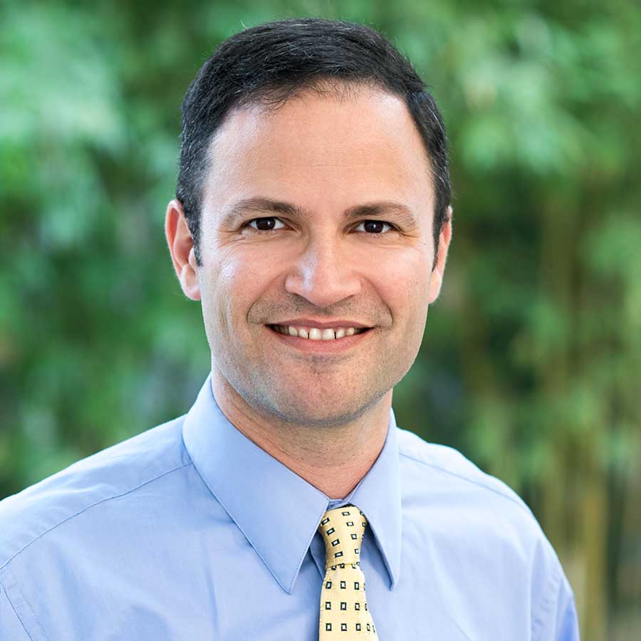
Jeffrey L. Goldberg, MD, PhD
Jeffrey L. Goldberg, MD, PhD is Professor and Chair of Ophthalmology at the Byers Eye Institute at Stanford University School of Medicine. Dr. Goldberg is a scientific advisor for the Catalyst for a Cure Vision Restoration Initiative (CFC3).